Amalgam tattoo or melanoma information
Home » information » Amalgam tattoo or melanoma informationYour Amalgam tattoo or melanoma images are available in this site. Amalgam tattoo or melanoma are a topic that is being searched for and liked by netizens today. You can Find and Download the Amalgam tattoo or melanoma files here. Get all free photos.
If you’re searching for amalgam tattoo or melanoma images information connected with to the amalgam tattoo or melanoma interest, you have pay a visit to the right site. Our website frequently provides you with hints for seeking the maximum quality video and image content, please kindly search and locate more enlightening video content and images that fit your interests.
Amalgam Tattoo Or Melanoma. A biopsy of the interdental palatal mucosa in the area of tooth 23 however revealed invasive atypical pleomorphic melanocytes with a positive expression for human melanoma black 45 consistent with mucosal melanoma. Amalgam tattoos develop as an amalgam deposit often as a result of continuous contact between an amalgam filling and the gingiva or amalgam fragments embedded in the oral tissue during dental filling or surgical operations. 21175766 PubMed - indexed for MEDLINE Publication Types. Amalgam is a common material used for dental fillings and contains silver tin mercury and other metals.
 Amalgam Tattoo In A Patient With Prior History Of Melanoma A Case Report Semantic Scholar From semanticscholar.org
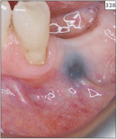
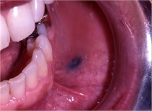
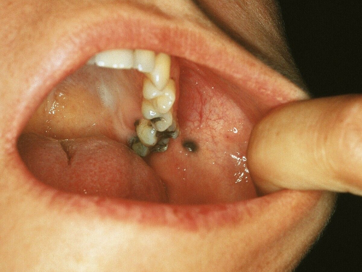
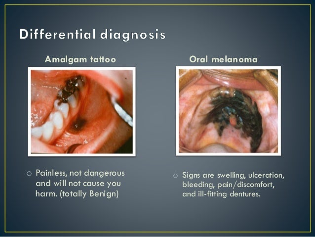
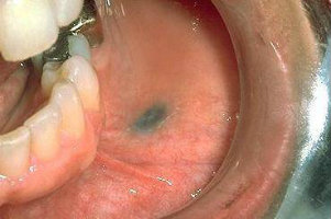
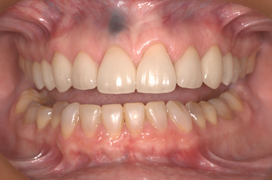
An amalgam tattoo refers to a deposit of particles in the tissue of your mouth usually from a dental procedure. When the amalgam is removed with a high-speed dental handpiece amalgam particles can be embedded or traumatically implanted in. The amalgam tattoo as shown below is a frequent finding in persons who have had dental amalgam restorations ie silver fillings in teeth. A biopsy of the interdental palatal mucosa in the area of tooth 23 however revealed invasive atypical pleomorphic melanocytes with a positive expression for human melanoma black 45 consistent with mucosal melanoma. Amalgam tattoos develop as an amalgam deposit often as a result of continuous contact between an amalgam filling and the gingiva or amalgam fragments embedded in the oral tissue during dental filling or surgical operations. Amalgam tattoo of the oral mucosa mimics malignant melanoma.
Amalgam consists of an alloy of liquid mercury with varying amounts of silver tin copper and zinc.
Mucosal melanoma is very rare but more advanced stages. More rarely palate and floor of the mouth. Amalgam tattoos are common oral pigmented lesions that clinically present as isolated blue grey or black macules on the gingivae the buccal and alveolar mucosae the palate andor the tongue. Images in Clinical Medicine from The New England Journal of Medicine Oral Amalgam Tattoo Mimicking Melanoma. Amalgam tattoo of the oral mucosa mimics malignant melanoma. Amalgam consists of an alloy of liquid mercury with varying amounts of silver tin copper and zinc.
 Source: dentagama.com
If you smoke you are far more likely to experience oral cancer than someone who does not. Sometimes fragments of amalgam restorations are broken off during extraction and embed in the adjacent soft tissue. Usually related to lower ridge or buccal vestibule. Amalgam tattoos are common oral pigmented lesions that clinically present as isolated blue grey or black macules on the gingivae the buccal and alveolar mucosae the palate andor the tongue. A biopsy of the interdental palatal mucosa in the area of tooth 23 however revealed invasive atypical pleomorphic melanocytes with a positive expression for human melanoma black 45 consistent with mucosal melanoma.
 Source: semanticscholar.org
A clinical diagnosis of amalgam tattoo was considered given the proximity of these pigmentations with dental restorations. By contrast amalgam tattoos formerly called localized argyria is one of the most frequent causes of exogenous pigmentation in the oral mucosa and is not uncommon to presentastwoormorelesions asseeninthepresent case. Amalgam tattoo of the oral mucosa mimics malignant melanoma. Amalgam tattoo is a common localized area of blue gray or black pigmentation caused by amalgam that has been embedded into the oral tissues during dental procedures. Amalgam is a common material used for dental fillings and contains silver tin mercury and other metals.
 Source: semanticscholar.org
Usually related to lower ridge or buccal vestibule. This deposit looks like a flat blue gray or. Another important difference between amalgam tattoos and mucosal melanoma is that although anyone with an amalgam filling can have an amalgam tattoo the vast majority of mucosal melanoma occur in chronic long-term tobacco users. By contrast amalgam tattoos formerly called localized argyria is one of the most frequent causes of exogenous pigmentation in the oral mucosa and is not uncommon to present as two or more lesions 5 13 15 as seen in the present case. Amalgam tattoos are common oral pigmented lesions that clinically present as isolated blue grey or black macules on the gingivae the buccal and alveolar mucosae the palate andor the tongue.
 Source: scialert.net
Amalgam consists of an alloy of liquid mercury with varying amounts of silver tin copper and zinc. By contrast amalgam tattoos formerly called localized argyria is one of the most frequent causes of exogenous pigmentation in the oral mucosa and is not uncommon to presentastwoormorelesions asseeninthepresent case. Amalgam is a common material used for dental fillings and contains silver tin mercury and other metals. Usually related to lower ridge or buccal vestibule. More rarely palate and floor of the mouth.
 Source: pocketdentistry.com
Another important difference between amalgam tattoos and mucosal melanoma is that although anyone with an amalgam filling can have an amalgam tattoo the vast majority of mucosal melanoma occur in chronic long-term tobacco users. They are due to deposition of a mixture of silver tin mercury copper and zinc which are components of an amalgam filling into the oral soft tissues. Amalgam Tattoo Focal argyrosis Clinical features Black or bluish-black usually solitary nonelevated small pigmented area beneath normal mucosa. Another important difference between amalgam tattoos and mucosal melanoma is that although anyone with an amalgam filling can have an amalgam tattoo the vast majority of mucosal melanoma occur in chronic long-term tobacco users. Images in Clinical Medicine from The New England Journal of Medicine Oral Amalgam Tattoo Mimicking Melanoma.
 Source: dentagama.com
By contrast amalgam tattoos formerly called localized argyria is one of the most frequent causes of exogenous pigmentation in the oral mucosa and is not uncommon to present as two or more lesions 5 13 15 as seen in the present case. Mucosal melanoma is very rare but more advanced stages. Sometimes fragments of amalgam restorations are broken off during extraction and embed in the adjacent soft tissue. The amalgam tattoo as shown below is a frequent finding in persons who have had dental amalgam restorations ie silver fillings in teeth. Further a galvanic element can form if various.
 Source: semanticscholar.org
A clinical diagnosis of amalgam tattoo was considered given the proximity of these pigmentations with dental restorations. More rarely palate and floor of the mouth. This deposit looks like a flat blue gray or. An amalgam tattoo refers to a deposit of particles in the tissue of your mouth usually from a dental procedure. When the amalgam is removed with a high-speed dental handpiece amalgam particles can be embedded or traumatically implanted in.
 Source: altmeyers.org
Most amalgam tattoos are due to dental treatments when metal particles accidentally deposit in open oral wounds or are dashed like a shrapnel into the oral mucosa during tooth preparation procedures. Amalgam consists of an alloy of liquid mercury with varying amounts of silver tin copper and zinc. If you smoke you are far more likely to experience oral cancer than someone who does not. Amalgam is a common material used for dental fillings and contains silver tin mercury and other metals. Another important difference between amalgam tattoos and mucosal melanoma is that although anyone with an amalgam filling can have an amalgam tattoo the vast majority of mucosal melanoma occur in chronic long-term tobacco users.
 Source: exodontia.info
Amalgam Tattoo Focal argyrosis Clinical features Black or bluish-black usually solitary nonelevated small pigmented area beneath normal mucosa. It is asymptomatic and may rarely be radiopaque. 21175766 PubMed - indexed for MEDLINE Publication Types. Amalgam is a common material used for dental fillings and contains silver tin mercury and other metals. More rarely palate and floor of the mouth.
 Source: onlinelibrary.wiley.com
The amalgam tattoo as shown below is a frequent finding in persons who have had dental amalgam restorations ie silver fillings in teeth. By contrast amalgam tattoos formerly called localized argyria is one of the most frequent causes of exogenous pigmentation in the oral mucosa and is not uncommon to presentastwoormorelesions asseeninthepresent case. A clinical diagnosis of amalgam tattoo was considered given the proximity of these pigmentations with dental restorations. The amalgam tattoo as shown below is a frequent finding in persons who have had dental amalgam restorations ie silver fillings in teeth. Most amalgam tattoos are due to dental treatments when metal particles accidentally deposit in open oral wounds or are dashed like a shrapnel into the oral mucosa during tooth preparation procedures.
 Source: slideshare.net
Amalgam tattoos develop as an amalgam deposit often as a result of continuous contact between an amalgam filling and the gingiva or amalgam fragments embedded in the oral tissue during dental filling or surgical operations. Amalgam tattoos can resemble spots of mucosal melanoma a type of skin cancer of internal membranes. When the amalgam is removed with a high-speed dental handpiece amalgam particles can be embedded or traumatically implanted in. If you smoke you are far more likely to experience oral cancer than someone who does not. Sometimes fragments of amalgam restorations are broken off during extraction and embed in the adjacent soft tissue.
 Source: just-teeth.co.nz
Amalgam Tattoo Focal argyrosis Clinical features Black or bluish-black usually solitary nonelevated small pigmented area beneath normal mucosa. The amalgam tattoo as shown below is a frequent finding in persons who have had dental amalgam restorations ie silver fillings in teeth. It is asymptomatic and may rarely be radiopaque. Amalgam tattoo is a common localized area of blue gray or black pigmentation caused by amalgam that has been embedded into the oral tissues during dental procedures. Most amalgam tattoos are due to dental treatments when metal particles accidentally deposit in open oral wounds or are dashed like a shrapnel into the oral mucosa during tooth preparation procedures.
 Source: sciencedirect.com
The amalgam tattoo as shown below is a frequent finding in persons who have had dental amalgam restorations ie silver fillings in teeth. Amalgam is a common material used for dental fillings and contains silver tin mercury and other metals. Amalgam tattoos can resemble spots of mucosal melanoma a type of skin cancer of internal membranes. By contrast amalgam tattoos formerly called localized argyria is one of the most frequent causes of exogenous pigmentation in the oral mucosa and is not uncommon to present as two or more lesions 5 13 15 as seen in the present case. The amalgam tattoo as shown below is a frequent finding in persons who have had dental amalgam restorations ie silver fillings in teeth.
 Source: hindawi.com
This deposit looks like a flat blue gray or. Amalgam is a common material used for dental fillings and contains silver tin mercury and other metals. If you smoke you are far more likely to experience oral cancer than someone who does not. Sometimes fragments of amalgam restorations are broken off during extraction and embed in the adjacent soft tissue. Most amalgam tattoos are due to dental treatments when metal particles accidentally deposit in open oral wounds or are dashed like a shrapnel into the oral mucosa during tooth preparation procedures.
 Source: healthline.com
Most amalgam tattoos are due to dental treatments when metal particles accidentally deposit in open oral wounds or are dashed like a shrapnel into the oral mucosa during tooth preparation procedures. Amalgam tattoo is a common localized area of blue gray or black pigmentation caused by amalgam that has been embedded into the oral tissues during dental procedures. Amalgam consists of an alloy of liquid mercury with varying amounts of silver tin copper and zinc. Amalgam tattoos can resemble spots of mucosal melanoma a type of skin cancer of internal membranes. When the amalgam is removed with a high-speed dental handpiece amalgam particles can be embedded or traumatically implanted in.
 Source: exodontia.info
Amalgam consists of an alloy of liquid mercury with varying amounts of silver tin copper and zinc. By contrast amalgam tattoos formerly called localized argyria is one of the most frequent causes of exogenous pigmentation in the oral mucosa and is not uncommon to present as two or more lesions 5 13 15 as seen in the present case. A biopsy of the interdental palatal mucosa in the area of tooth 23 however revealed invasive atypical pleomorphic melanocytes with a positive expression for human melanoma black 45 consistent with mucosal melanoma. Further a galvanic element can form if various. Another important difference between amalgam tattoos and mucosal melanoma is that although anyone with an amalgam filling can have an amalgam tattoo the vast majority of mucosal melanoma occur in chronic long-term tobacco users.
 Source: hindawi.com
Amalgam tattoos can resemble spots of mucosal melanoma a type of skin cancer of internal membranes. A biopsy of the interdental palatal mucosa in the area of tooth 23 however revealed invasive atypical pleomorphic melanocytes with a positive expression for human melanoma black 45 consistent with mucosal melanoma. The amalgam tattoo as shown below is a frequent finding in persons who have had dental amalgam restorations ie silver fillings in teeth. Usually related to lower ridge or buccal vestibule. Amalgam tattoo is a common localized area of blue gray or black pigmentation caused by amalgam that has been embedded into the oral tissues during dental procedures.
 Source: dentagama.com
It is asymptomatic and may rarely be radiopaque. A clinical diagnosis of amalgam tattoo was considered given the proximity of these pigmentations with dental restorations. This deposit looks like a flat blue gray or. An amalgam tattoo refers to a deposit of particles in the tissue of your mouth usually from a dental procedure. By contrast amalgam tattoos formerly called localized argyria is one of the most frequent causes of exogenous pigmentation in the oral mucosa and is not uncommon to presentastwoormorelesions asseeninthepresent case.
This site is an open community for users to do sharing their favorite wallpapers on the internet, all images or pictures in this website are for personal wallpaper use only, it is stricly prohibited to use this wallpaper for commercial purposes, if you are the author and find this image is shared without your permission, please kindly raise a DMCA report to Us.
If you find this site good, please support us by sharing this posts to your own social media accounts like Facebook, Instagram and so on or you can also save this blog page with the title amalgam tattoo or melanoma by using Ctrl + D for devices a laptop with a Windows operating system or Command + D for laptops with an Apple operating system. If you use a smartphone, you can also use the drawer menu of the browser you are using. Whether it’s a Windows, Mac, iOS or Android operating system, you will still be able to bookmark this website.
Category
Related By Category
- Carbs in baked beans ideas
- Albuterol and blood sugar ideas
- How long does leg waxing last information
- Scrotox before and after information
- Tanfast information
- Is mane and tail safe for color treated hair information
- Coconut oil after microneedling ideas
- Paleo psoriasis ideas
- Erythema marginatum vs multiforme ideas
- Self shoulder adjustment information