The radiographic imaging of blood vessels of the eye with fluorescing dye is ideas
Home » health » The radiographic imaging of blood vessels of the eye with fluorescing dye is ideasYour The radiographic imaging of blood vessels of the eye with fluorescing dye is images are available in this site. The radiographic imaging of blood vessels of the eye with fluorescing dye is are a topic that is being searched for and liked by netizens now. You can Get the The radiographic imaging of blood vessels of the eye with fluorescing dye is files here. Download all free images.
If you’re searching for the radiographic imaging of blood vessels of the eye with fluorescing dye is pictures information related to the the radiographic imaging of blood vessels of the eye with fluorescing dye is interest, you have pay a visit to the ideal site. Our site always provides you with suggestions for refferencing the highest quality video and picture content, please kindly surf and find more enlightening video content and graphics that match your interests.
The Radiographic Imaging Of Blood Vessels Of The Eye With Fluorescing Dye Is. Radiographic imaging of blood vessels of the eye with fluorescing dye. The first surge of blood termed an E-wave is associated with atrial diastole and results from the pressure gradient between the venous system and ventricle whereas the second surge of blood termed an A-wave is a result of energy imparted to the blood. Instrument used for visual examination of the eye. Trial data with this.
 Orbital Blowout Fracture Teardrop Sign Blowout Herniated Ocular From za.pinterest.com
This is useful for both diagnosis and therapy and occurs in. Exercise 21 Pronunciation Exercise Exercise 22 1 retinal photocoagulation 2 from MEDICAL TE 101 at Champlain College. Radiographic imaging of blood vessels of the eye with fluorescing dye keratometer. Echo-Doppler imaging in a variety of anesthetized or physically restrained teleost and elasmobranch fishes shows biphasic filling of the ventricle. Trial data with this. Laser speckle imaging is a non-invasive technique that has been used for assessing tissue perfusion in multiple settings including intraoperatively during breast reconstruction and to assess gastric blood flow after esophagectomy59 60 The technique is designed to detect movement of red blood cells based on laser light formation and it is able to detect perfusion to the level of the dermis.
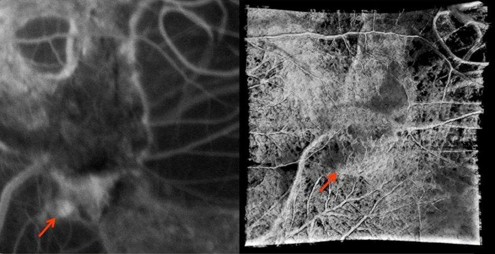
The radiographic imaging of blood vessels of the eye with fluorescing dyeis Afluorescein angiography.
Instrument used to measure the cornea. Radiographic imaging of blood vessels of the eye with fluorescing dye keratometer instrument used to measure curvature of the cornea used for fitting contact lenses. Radiographic imaging of blood vessels of the eye with fluorescing dye keratometer. Radiographic imaging of a blood vessel c. This preview shows page 24 - 27 out of 38 pagespreview shows page 24 - 27 out of 38 pages. Radiographic imaging of blood vessels of the eye with fluorescing dye keratometer.
 Source: healio.com
Transparent anterior part of the eyeball covering the iris pupil and anterior chamber that functions to refract bend light to focus a visual image choroid middle layer of the eye which is interlaced with many blood vessels that supply nutrients to the eye. The first surge of blood termed an E-wave is associated with atrial diastole and results from the pressure gradient between the venous system and ventricle whereas the second surge of blood termed an A-wave is a result of energy imparted to the blood. Radiographic imaging of a blood vessel c. Exercise 21 Pronunciation Exercise Exercise 22 1 retinal photocoagulation 2 from MEDICAL TE 101 at Champlain College. The radiographic imaging of blood vessels of the eye with fluorescing dyeis Afluorescein angiography.
 Source: mdpi.com
The obstetrician diagnosed her condition as A congenital defect in the vertebral column caused by the failure of the vertebral arch to close is called The term that means congenital herniation at the umbilicus is called The combining form meaning umbilicus navel is The word part that means bear give birth to labor childbirth is The term nulligravida refers to a woman who The term. Fluoroscopy is an imaging technique that uses X-rays to obtain real-time moving images of the interior of an object. Radiographic image of an artery. In its primary application of medical imaging a fluoroscope allows a physician to see the internal structure and function of a patient so that the pumping action of the heart or the motion of swallowing for example can be watched. Trial data with this.
 Source: researchgate.net
Instrument used to measure the curvature of the cornea used for fitting contact lenses ophthalmoscope. In its primary application of medical imaging a fluoroscope allows a physician to see the internal structure and function of a patient so that the pumping action of the heart or the motion of swallowing for example can be watched. Radiographic imaging of blood vessels of the eye with fluorescing dye keratometer. This preview shows page 24 - 27 out of 38 pagespreview shows page 24 - 27 out of 38 pages. Visual examination of a blood vessel d.
 Source: pinterest.com
Visual examination of a blood vessel d. In its primary application of medical imaging a fluoroscope allows a physician to see the internal structure and function of a patient so that the pumping action of the heart or the motion of swallowing for example can be watched. Radiographic image of an artery. Radiographic imaging of a blood vessel c. Fluoroscopy is an imaging technique that uses X-rays to obtain real-time moving images of the interior of an object.
 Source: pinterest.com
This preview shows page 24 - 27 out of 38 pagespreview shows page 24 - 27 out of 38 pages. Radiographic imaging of blood vessels of the eye with fluorescing dye keratometer instrument used to measure curvature of the cornea used for fitting contact lenses. Excision within the artery of plaque from the arterial wall is called. Instrument used to measure the cornea. In accordance with the present invention there are provided compositions useful for the in vivo delivery of a biologic wherein the biologic is associated with a polymeric shell formulated from a bio.
 Source: neurologic.theclinics.com
The radiographic imaging of blood vessels of the eye with fluorescing dyeis Afluorescein angiography. This is useful for both diagnosis and therapy and occurs in. Radiographic imaging of blood vessels of the eye with fluorescing dye keratometer instrument used to measure curvature of the cornea used for fitting contact lenses. The first surge of blood termed an E-wave is associated with atrial diastole and results from the pressure gradient between the venous system and ventricle whereas the second surge of blood termed an A-wave is a result of energy imparted to the blood. Laser speckle imaging is a non-invasive technique that has been used for assessing tissue perfusion in multiple settings including intraoperatively during breast reconstruction and to assess gastric blood flow after esophagectomy59 60 The technique is designed to detect movement of red blood cells based on laser light formation and it is able to detect perfusion to the level of the dermis.
 Source: neurologic.theclinics.com
Radiographic imaging of blood vessels of the eye with fluorescing dye keratometer. This preview shows page 24 - 27 out of 38 pagespreview shows page 24 - 27 out of 38 pages. Laser speckle imaging is a non-invasive technique that has been used for assessing tissue perfusion in multiple settings including intraoperatively during breast reconstruction and to assess gastric blood flow after esophagectomy59 60 The technique is designed to detect movement of red blood cells based on laser light formation and it is able to detect perfusion to the level of the dermis. Radiographic imaging of blood vessels of the eye with fluorescing dye keratometer. Radiographic imaging of blood vessels of the eye with fluorescing dye keratometer instrument used to measure curvature of the cornea used for fitting contact lenses.
 Source: nejm.org
Department of Diagnostic Radiology MD Anderson Cancer Center USA. Radiographic imaging of blood vessels of the eye with fluorescing dye keratometer. Laser speckle imaging is a non-invasive technique that has been used for assessing tissue perfusion in multiple settings including intraoperatively during breast reconstruction and to assess gastric blood flow after esophagectomy59 60 The technique is designed to detect movement of red blood cells based on laser light formation and it is able to detect perfusion to the level of the dermis. Fluoroscopy is an application of projectional radiography that allows real-time observation of the internal structures of a patient and is used predominantly in gastrointestinal imaging interventional radiology and musculoskeletal radiology. Visual examination of a blood vessel d.
Source:
Radiographic imaging of blood vessels of the eye with fluorescing dye keratometer. Visual examination of a blood vessel d. Fluoroscopy is an application of projectional radiography that allows real-time observation of the internal structures of a patient and is used predominantly in gastrointestinal imaging interventional radiology and musculoskeletal radiology. This preview shows page 24 - 27 out of 38 pagespreview shows page 24 - 27 out of 38 pages. In accordance with the present invention there are provided compositions useful for the in vivo delivery of a biologic wherein the biologic is associated with a polymeric shell formulated from a bio.
 Source: pinterest.com
In its primary application of medical imaging a fluoroscope allows a physician to see the internal structure and function of a patient so that the pumping action of the heart or the motion of swallowing for example can be watched. Visual examination of a blood vessel d. This preview shows page 24 - 27 out of 38 pagespreview shows page 24 - 27 out of 38 pages. Department of Diagnostic Radiology MD Anderson Cancer Center USA. The radiographic imaging of blood vessels of the eye with fluorescing dye is Select one.
 Source: pinterest.com
The radiographic imaging of blood vessels of the eye with fluorescing dyeis Afluorescein angiography. The radiographic imaging of blood vessels of the eye with fluorescing dye is Select one. Radiographic imaging of blood vessels of the eye with fluorescing dye. Transparent anterior part of the eyeball covering the iris pupil and anterior chamber that functions to refract bend light to focus a visual image choroid middle layer of the eye which is interlaced with many blood vessels that supply nutrients to the eye. The first surge of blood termed an E-wave is associated with atrial diastole and results from the pressure gradient between the venous system and ventricle whereas the second surge of blood termed an A-wave is a result of energy imparted to the blood.
 Source: researchgate.net
Radiographic imaging of blood vessels of the eye with fluorescing dye keratometer. The first surge of blood termed an E-wave is associated with atrial diastole and results from the pressure gradient between the venous system and ventricle whereas the second surge of blood termed an A-wave is a result of energy imparted to the blood. The obstetrician diagnosed her condition as A congenital defect in the vertebral column caused by the failure of the vertebral arch to close is called The term that means congenital herniation at the umbilicus is called The combining form meaning umbilicus navel is The word part that means bear give birth to labor childbirth is The term nulligravida refers to a woman who The term. The radiographic imaging of blood vessels of the eye with fluorescing dye is Select one. Radiographic imaging of blood vessels of the eye with fluorescing dye keratometer instrument used to measure the curvature of the cornea used for fitting contact lenses.
 Source: in.pinterest.com
The first surge of blood termed an E-wave is associated with atrial diastole and results from the pressure gradient between the venous system and ventricle whereas the second surge of blood termed an A-wave is a result of energy imparted to the blood. Laser speckle imaging is a non-invasive technique that has been used for assessing tissue perfusion in multiple settings including intraoperatively during breast reconstruction and to assess gastric blood flow after esophagectomy59 60 The technique is designed to detect movement of red blood cells based on laser light formation and it is able to detect perfusion to the level of the dermis. The radiographic imaging of blood vessels of the eye with fluorescing dyeis Afluorescein angiography. Instrument used to measure the curvature of the cornea used for fitting contact lenses ophthalmoscope. Radiographic imaging of blood vessels of the eye with fluorescing dye keratometer instrument used to measure curvature of the cornea used for fitting contact lenses.
 Source: delhieyecentre.in
Radiographic imaging of a blood vessel c. Radiographic imaging of blood vessels of the eye with fluorescing dye keratometer. Instrument used to measure the cornea. Trial data with this. Near-infrared NIR fluorescence imaging is usually used in concert with molecularly targeted contrast agents that not only provide enhanced contrast but also more importantly reveal specific molecular events associated with cancer formation and progression.
 Source: pinterest.com
Exercise 21 Pronunciation Exercise Exercise 22 1 retinal photocoagulation 2 from MEDICAL TE 101 at Champlain College. Radiographic image of an artery. Visual examination of a blood vessel d. Excision within the artery of plaque from the arterial wall is called. This is useful for both diagnosis and therapy and occurs in.
 Source: tbrhsc.net
Fluoroscopy is an application of projectional radiography that allows real-time observation of the internal structures of a patient and is used predominantly in gastrointestinal imaging interventional radiology and musculoskeletal radiology. Radiographic imaging of blood vessels of the eye with fluorescing dye keratometer instrument used to measure the curvature of the cornea used for fitting contact lenses. Instrument used to measure the cornea. Echo-Doppler imaging in a variety of anesthetized or physically restrained teleost and elasmobranch fishes shows biphasic filling of the ventricle. This preview shows page 24 - 27 out of 38 pagespreview shows page 24 - 27 out of 38 pages.
 Source: co.pinterest.com
The obstetrician diagnosed her condition as A congenital defect in the vertebral column caused by the failure of the vertebral arch to close is called The term that means congenital herniation at the umbilicus is called The combining form meaning umbilicus navel is The word part that means bear give birth to labor childbirth is The term nulligravida refers to a woman who The term. Instrument used for visual examination of the interior of the eye. Radiographic imaging of a blood vessel c. Transparent anterior part of the eyeball covering the iris pupil and anterior chamber that functions to refract bend light to focus a visual image choroid middle layer of the eye which is interlaced with many blood vessels that supply nutrients to the eye. Instrument used for visual examination of the eye.
 Source: cureus.com
Radiographic imaging of blood vessels of the eye with fluorescing dye keratometer. Visual examination of a blood vessel d. Echo-Doppler imaging in a variety of anesthetized or physically restrained teleost and elasmobranch fishes shows biphasic filling of the ventricle. Laser speckle imaging is a non-invasive technique that has been used for assessing tissue perfusion in multiple settings including intraoperatively during breast reconstruction and to assess gastric blood flow after esophagectomy59 60 The technique is designed to detect movement of red blood cells based on laser light formation and it is able to detect perfusion to the level of the dermis. Exercise 21 Pronunciation Exercise Exercise 22 1 retinal photocoagulation 2 from MEDICAL TE 101 at Champlain College.
This site is an open community for users to share their favorite wallpapers on the internet, all images or pictures in this website are for personal wallpaper use only, it is stricly prohibited to use this wallpaper for commercial purposes, if you are the author and find this image is shared without your permission, please kindly raise a DMCA report to Us.
If you find this site convienient, please support us by sharing this posts to your favorite social media accounts like Facebook, Instagram and so on or you can also bookmark this blog page with the title the radiographic imaging of blood vessels of the eye with fluorescing dye is by using Ctrl + D for devices a laptop with a Windows operating system or Command + D for laptops with an Apple operating system. If you use a smartphone, you can also use the drawer menu of the browser you are using. Whether it’s a Windows, Mac, iOS or Android operating system, you will still be able to bookmark this website.
Category
Related By Category
- Banzai bowl nutrition facts ideas
- Fish without bones ideas
- Dennie morgan lines information
- Can you eat after a filling information
- Zofran breastfeeding ideas
- Essential oils for fever ideas
- Torus palatinus sore information
- Nursing night light information
- Feels like something crawling in my eye information
- Choledochlithiasis ideas